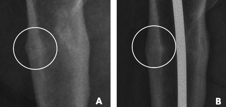
Figure 7.
Endocortical callus formation A, There are some callus on fracture site, but we can see just blurred image at preoperatively. B, For 4 weeks since the PEIN, the radiograph shows ongoing bone healing process, and we can see endocortical callus at the medial side of fracture line. Callus is becoming more radiopaque as a cortical bone’s signal and “completely connecting” the endocortical area.

