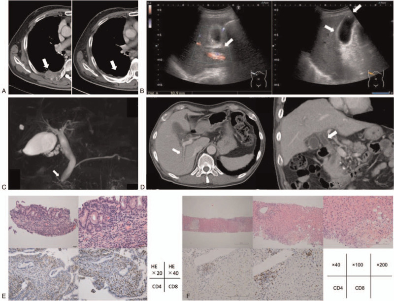
Figure 1.

(A) Computed tomography (CT). Right pleural metastasis was observed before the start of nivolumab administration as the second-line chemotherapy (arrow). After 11 courses, the patient was diagnosed as diabetes mellitus, but the CT scan showed shrinkage of the metastasis. No abnormal accumulation was observed on PET-CT so that we determined complete response. (B) Abdominal ultrasound. Dilatation of the common bile duct was 10.9 mm (arrow). Mild wall thickening of the gallbladder (arrow), and cholelith less than 10 mm (arrow) were observed. (C) Magnetic resonance cholangiopancreatography (MRCP). Mild dilatation of the common bile duct was shown (arrow). Multiple low signal areas were observed, and common bile duct stones or debris was suspected. (D) CT at the second time. Periportal collar (arrow), approximately 11.0 mm of dilatation of the common bile duct, and bile duct wall thickening (arrow) were observed. (E) Pathological findings of bile duct biopsy. [Upper left] HE 20× [upper right] HE 40×, [Lower left] CD4 staining 200× [lower middle] CD8 staining 200×. There was no sign of malignant finding due to lack of atypical bile duct epithelial. Infiltration by lymphocyte and plasma cells was observed in the interstitium. CD4-positive T cells and CD8-positive T cells were also observed. IgG4 was negative. (F) Pathological findings of liver biopsy. [Upper left] HE 40× [upper middle] HE 100× [upper right] HE 200×, [Lower left] CD4 staining 200× [lower middle] CD8 staining 200×. Mild fibrous expansion of portal canal was observed, and small bile duct was normal. Only a few inflammatory cell infiltration was observed. CD8-positive T cells were found. Bile infarct was observed only in the lower right area of slide, and no inflammatory cells were found around necrosis. No bile plug was formed. There was no sign of malignant finding. PET = positron emission tomography.
