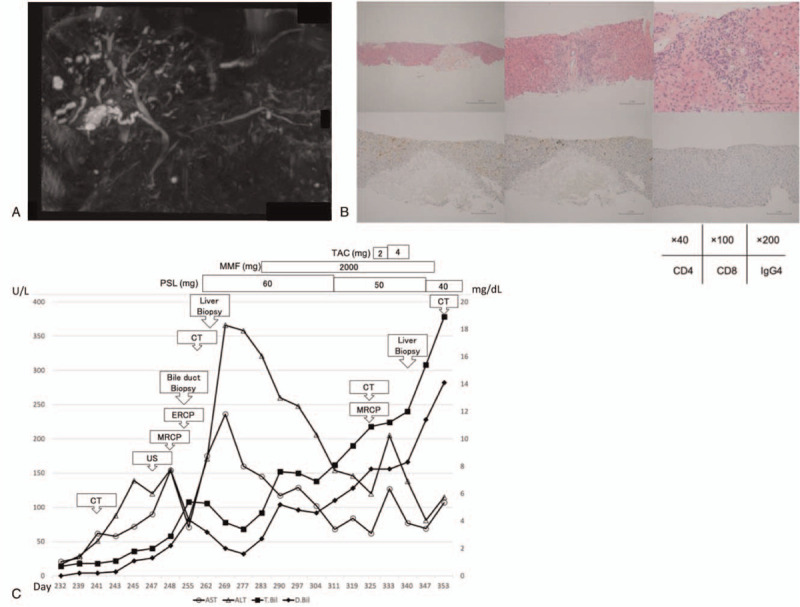
Figure 2.

(A) MRCP when bilirubin level was re-increased at the second time. Periportal intensity was found on T2-weighted image. Beaded constriction and dilatation of the peripheral intrahepatic bile duct were detected. No dilatation of the common bile duct was found, but there was wall thickening of extrahepatic bile duct. There was also circumferential wall thickening of bladder. (B) Pathological findings of liver biopsy at the second time, [Upper left] HE 40× [upper middle] HE 100× [upper right] HE 200×, [Lower left] CD4 staining 200× [lower middle] CD8 staining 200× [lower right] IgG4 staining 200×. In periportal area, inflammatory cell infiltration was scarce, and small bile duct was normal. Biliary hyperplasia with cholestasis was observed. Hepatocyte necrosis was sporadic, and normal cells were sharply defined. It was punched out necrosis. There was no migration of lymphocytes at the site of necrosis and were only a few CD8-positive T cells around. IgG was negative. (C) Summary of clinical course and biochemical examination. MRCP = magnetic resonance cholangiopancreatography.
