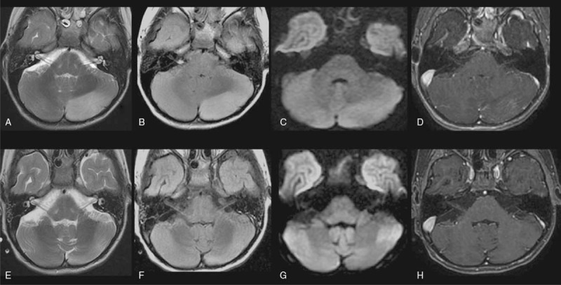
Figure 1.

Brain MRI of a pediatric patient with autoimmune anti-GABAA receptor encephalitis comorbid with an established B19 infection of the central nervous system at two timepoints. A–D. Images from the patient's first MRI scan, which occurred while he was being treated in the ICU for persistent seizures that evolved into focal SE with fluctuating consciousness. T2/FLAIR hyperintensity and mild expansion in the left cerebellar hemisphere, with some foci of contrast enhancement and an absence of restricted diffusion. E–H. Images from a follow-up scan performed almost a month later, likely showing residual post-inflammatory changes. Note that the previously observed T2/FLAIR hyperintensity and volume expansion were reduced and that there were no new lesions or areas of contrast uptake in the left cerebellar hemisphere.
