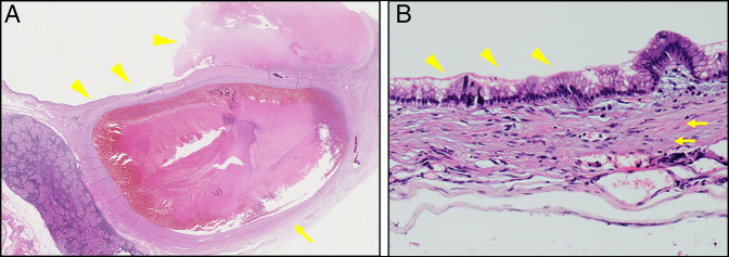
Figure 3.
Pathological image showing (A) splenic arterial aneurysm (arrow) and pancreatic cyst with hemorrhage (arrowhead) at low power field and (B) pancreatic cyst is consisted with ovarian-type stroma (arrow), and mucinous epithelium layer with papillary proliferation is lined up inside the lumen of the cyst (arrowhead) at high power field.

