
Fig. 1.
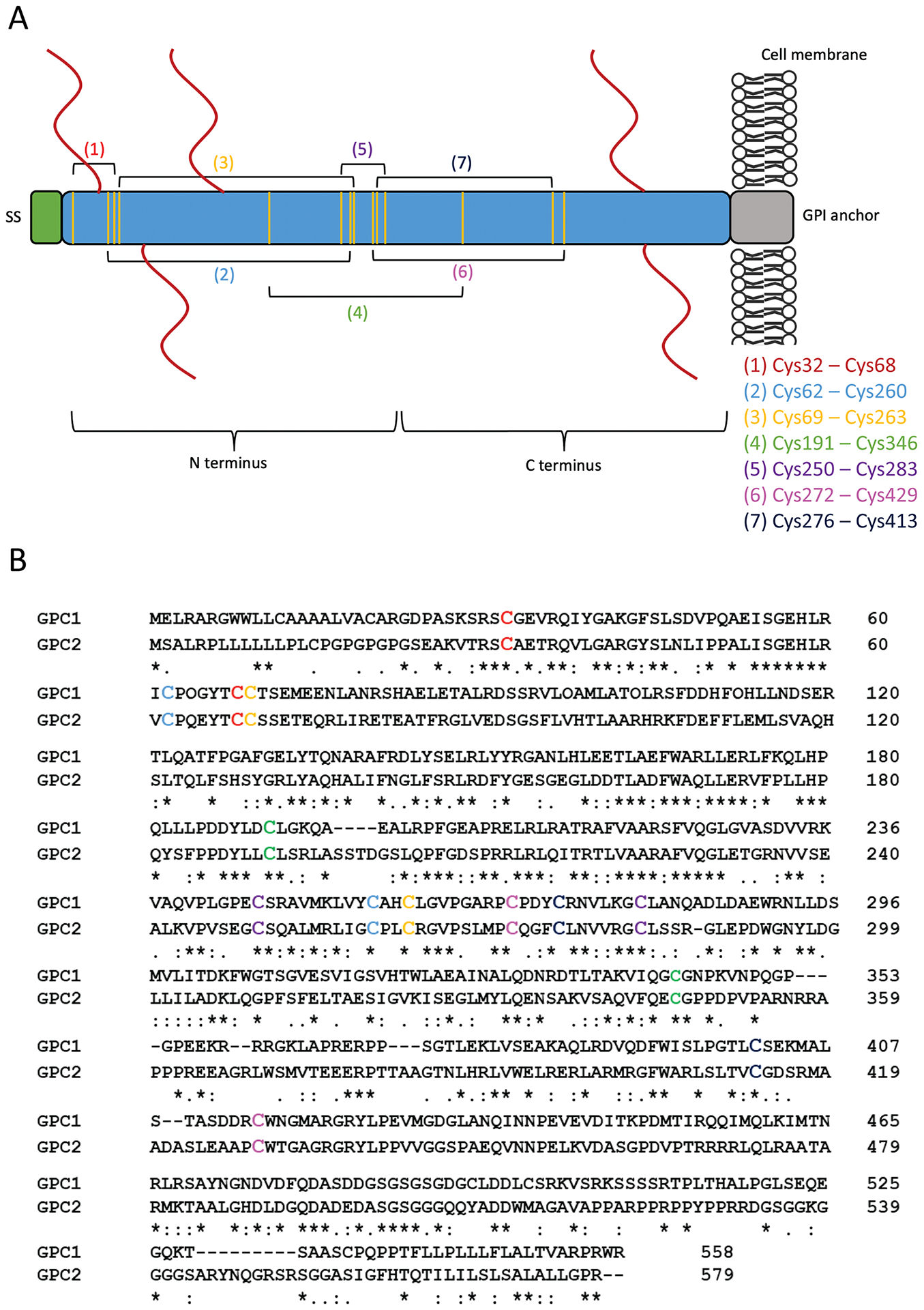
Structure and sequence of GPC2. (A) Schematic of the GPC2 protein. The C terminus is located at the cell surface; the N terminus is distal to the cell surface. The mature protein is 579 amino acids. The N-terminal signal peptide (SS) comprises the first 24 amino acids. The glycosylphosphatidylinositol (GPI) anchor attachment is located at G554. The predicted heparan sulfate (HS) chains are shown by red lines at S55, S92, S155, S500, and S502. The conserved cysteine residues are represented by the vertical yellow lines. Brackets drawn between yellow lines denote the 7 predicted disulfide bonds. The specific arrangement of the bonds is listed. (B) The sequence alignment of GPC1 and GPC2. The cysteine residues involved in the formation of disulfide bonds are shown in color corresponding to (A). The serine-glycine residues that serve as HS attachment sites on the GPC2 core protein are shown in black boxes.