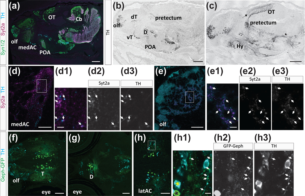
FIGURE 5.
Tyrosine hydroxylase (TH) expression and colocalization with Syt2a. (a–c) Sagittal view of a brain at 14 dpf, showing TH expression in distinct neuronal clusters throughout the brain. (a) TH (cyan), Syt2a (magenta), and Syn1/2 (green) expression in the brain. (b,c) TH is expressed in neurons in the olfactory bulb (olf), dorsal (dT), and ventral telencephalon (vT), diencephalon (D), preoptic area (POA), and pretectum. TH projections are located throughout the brain. An arrow marks labeling in the optic tectum (OT). (d–d3) Syt2a and TH colocalize at synapses in the AC. Arrows mark Syt2a (d2) and TH (d3) colabeled puncta. (e–e3) In the olfactory bulb we find many Syt2 (e2) and TH (e3) co-expressing puncta (arrows). (f–h3) Dorsal view of the forebrain showing TH expression (cyan) and GFP-Geph positive (green) specifically in BFCNsy321. (f) Expression in the olf and telencephalon (T). (g) More posterior view showing TH expression and GFP-Geph positive BFCNsy321 in the diencephalon (D). (h–h3) Higher magnification of the telencephalic region. A yellow and black asterisk marks TH only and GFP-Geph only neurons, respectively. Arrows point to TH and GFP-Geph colocalization on projections from BFCNsy321. Area shown in (h–h3) is marked with a box in (h) Scale bars are 200 μm in (a–c), 30 μm in (d,e), 70 μm in (f,g), 50 μm in (h), and 5 μm in (d1,e1,h1)

