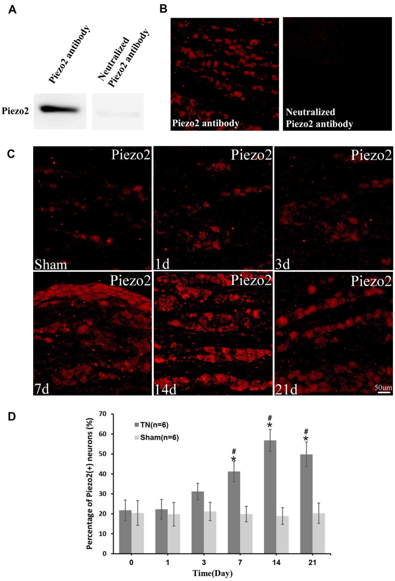
Figure 1.
Immunocytochemistry of Piezo2. The efficiency of Piezo2 antibody was confirmed by Western blotting and immunofluorescence (A, B). Immunocytochemistry images show the expression of Piezo2 in sham group and TN group at different time points (C). With quantification, the percentage of Piezo2 positive neuron at different time points is displayed, which reaches peak at day 14 (9.2±1.6% in TN group vs. 2.21±1.1% in sham group, p<0.05) (D). *p<0.05 vs. the sham group; # p<0.05 vs. the bassline.

