Figure 7.
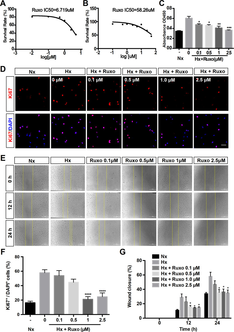
Ruxolitinib inhibits hypoxia-induced rPASMCs proliferation and migration. Nx = Normoxia group; Hx = Hypoxia group; Hx + Ruxo = Hypoxia plus Ruxolitinib group. (A, B) Determination of IC50 values. RPASMCs were incubated with ruxolitinib at different concentrations (0, 0.1, 0.5, 1 and 2.5 μM) for 24 h under hypoxia (A) and normoxia (B), viable cell number was determined by MTT assay. The results yielded IC50 values were 6.719 μM (A) and 58.26 μM (B), respectively. (C) CCK-8 assay for cell proliferation. RPASMCs were treated with indicated concentrations of ruxolitinib for 24 h, and the cell proliferation was determined by CCK-8 assay. (D, F) Immunofluorescence staining of Ki67(red) in rPASMCs of indicated groups. RPASMCs were treated with or without indicated concentrations of ruxolitinib for 24 h under hypoxic conditions, the untreated cells were treated as a normoxia control group. Cells nuclear were counterstained with DAPI (blue). (E, G) Wound scratch assay. RPASMCs were treated with ruxolitinib at a specified concentration in the presence or absence of oxygen for 12h and 24h, migration capabilities were represented by relative migration distances. Data are presented as mean ± SEM, *P < 0.05, **P < 0.01, ***P < 0.001, ****P < 0.0001.

