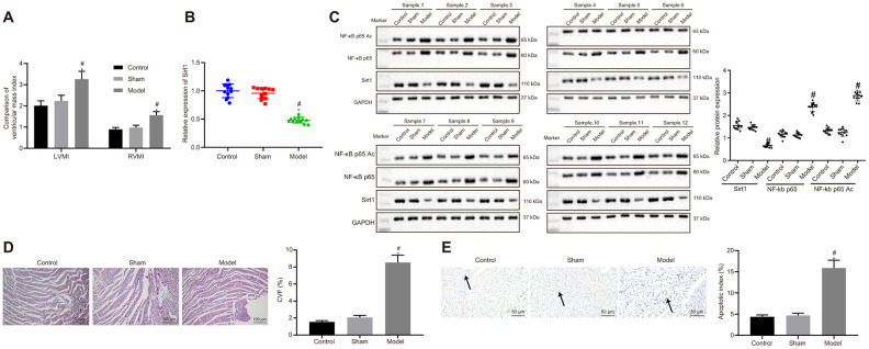
Figure 1.
Sirt1, NF-κB p65 expression and apoptosis in heart tissues of successfully induced HF rats. (A) Ventricular mass index; (B) Sirt1 mRNA expression, determined using RT-qPCR; (C) Sirt1, NF-κB p65, and NF-κB p65 Ac protein expression assessed by Western blot analysis; (D) Collagen volume faction determined by Masson’s trichrome staining (100 ×); (E) Apoptosis determined by TUNEL staining (200 ×); *p < 0.05 vs. control rats and # p < 0.05 vs. sham rats. Data were expressed as a mean ± standard deviation. Three or more groups were analyzed by one-way analysis of variance (ANOVA) and Tukey's post hoc test. N = 12.

