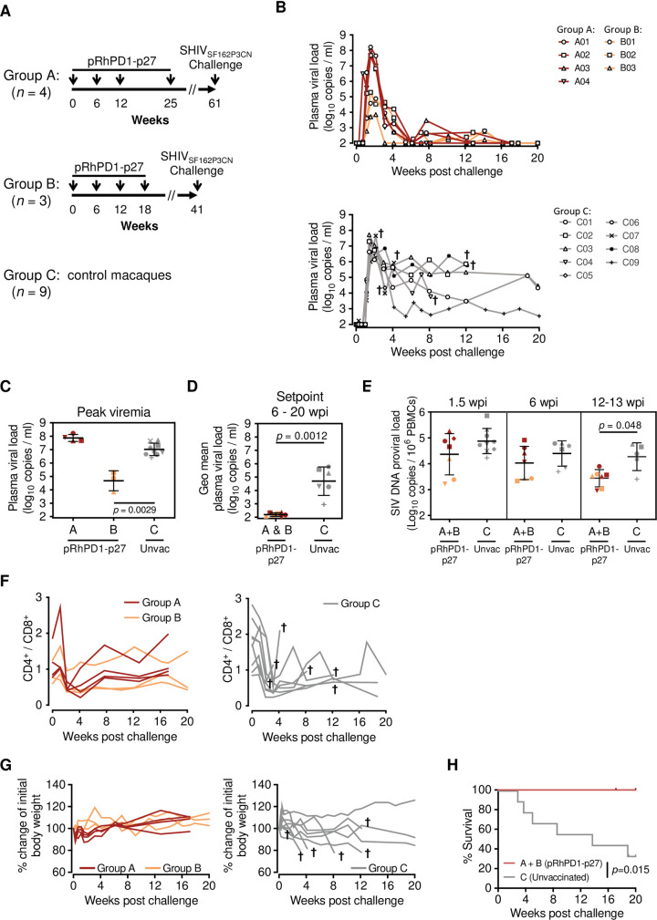
Fig 2. Viral suppression in rhesus macaques immunised with the PD1-based pRhPD1-p27 DNA vaccine after high dose pathogenic SHIVSF162P3CN challenge.
(A) Schematic of DNA vaccination in Chinese-origin rhesus macaques. Two studies with different vaccination durations were conducted. In both studies, Chinese-origin rhesus macaques were immunised with the PD1-based pRhPD1-p27 DNA vaccine via intramuscular injection with electroporation 4 times in 6- to 13-week intervals, followed by intravenous challenge with 5000 TCID50 of SHIVSF162P3CN at 23 or 36 weeks after last immunization. Unvaccinated animals were included as controls. (B) Plasma viral loads of the vaccinated (top) and unvaccinated macaques (below) after SHIV challenge. Animals that were euthanised were marked with †. Anti-CD8β antibody (clone CD8b255R1) was infused into the vaccinated macaques of Group A intravenously at 17 weeks post-challenge, after confirming pVL were below detection levels. Data of this treatment is presented in Fig 5D. (C) Comparison of the peak viremia and (D) geometric means of setpoint viremia determined from 6–20 weeks post-infection (wpi). (E) Proviral DNA loads in PBMC in vaccinated and unvaccinated macaques. Geometric means ± geometric SD are shown in (C)–(E). Significances of differences were determined by the Kruskal-Wallis test followed by the Dunn’s multiple comparisons test (C), or the two-tailed Wilcoxon rank sum test (D and E). (F) The CD4+/CD8+ T cell ratios in vaccinated rhesus macaques from Group A and B (left) or unvaccinated macaques (right) after SHIVSF162P3CN challenge. (G) Changes of body weight of the vaccinated (left) and unvaccinated macaques (right). Animals that were euthanised over the measurement period were marked with †. (H) Survival of vaccinated and unvaccinated macaques after SHIV challenge. The significance of difference was determined by log-rank test.

