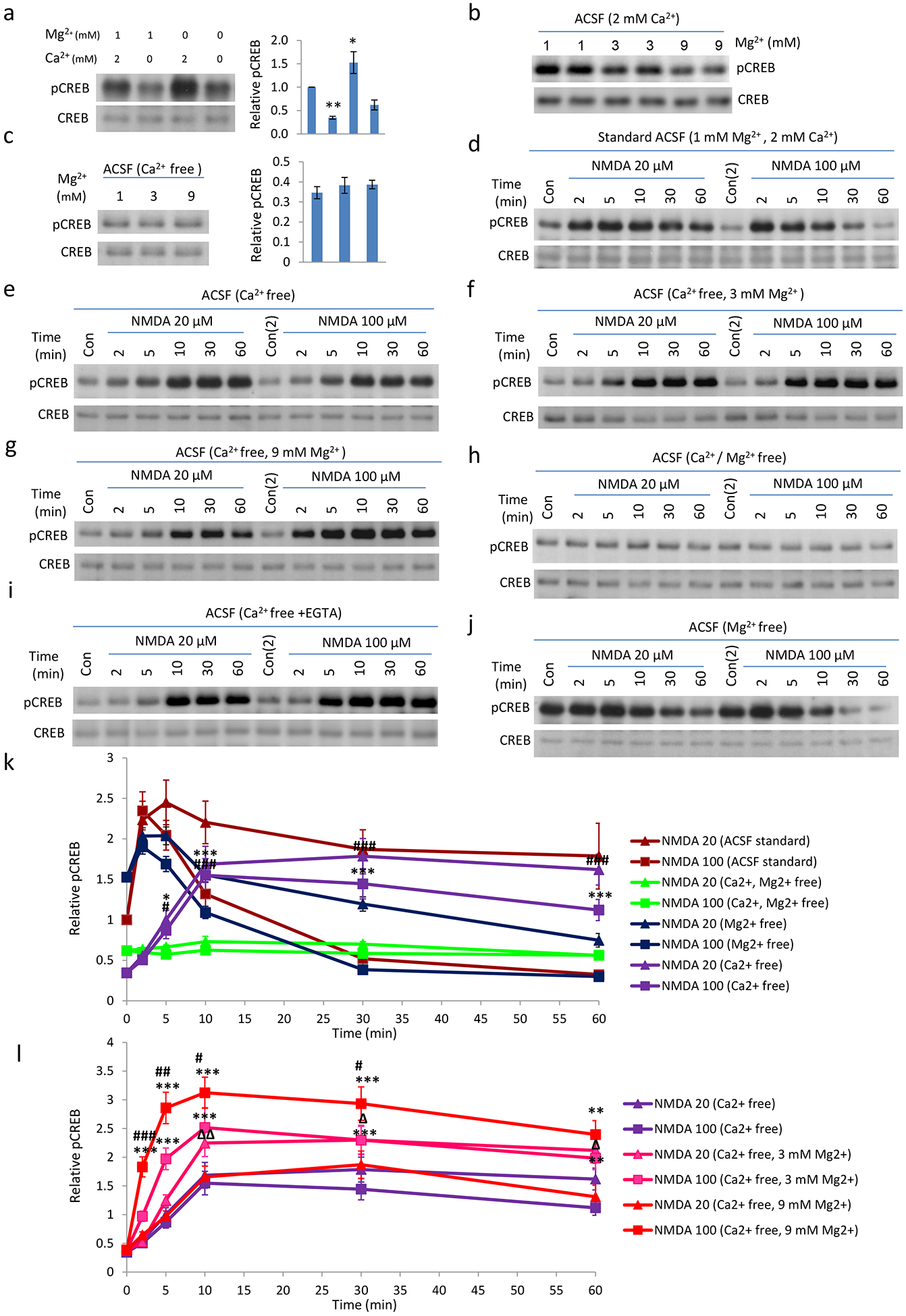
Fig. 3. Extracellular Mg2+ is responsible for the delayed CREB phosphorylation induced by NMDA in neurons incubated with Ca2+-free ACSF.

a. Basal pCREB level in Standard, Ca2+ free, Mg2+ free, and Ca2+ /Mg2+ free ACSF. Neurons were briefly washed with different ACSF (preconditioned in incubator for >1 hour) for 3 times, and then incubated in the same ACSF for 1 hour. Components of normal ACSF(mM: 125 NaCl; 2.5 KCl; 1 MgCl2; 2 CaCl2; 33 D-Glucose; 25 HEPES; pH7.3); 10 μM glycine was added freshly before experiments. b: Increasing magnesium concentration in ACSF decreases basal pCREB. c. Basal pCREB in Ca2+ free ACSF with different concentration of Mg2+. d-j. Time course of 20 or 100 μM NMDA in the regulation of CREB phosphorylation in neurons in different ACSF. Neurons were pre-incubated with different ACSF for 1 hour, before the pCREB time-course by NMDA was determined. Delayed CREB phosphorylation can be detected in Ca2+ free ACSF (e); Strong NMDAR stimulation (100 μM) cannot shut-off pCREB (e-g). Removal of Ca2+/Mg2+ abolished NMDA induced CREB phosphorylation (h); i: potential residual calcium in calcium free ACSF did not produce the delay pCREB by NMDA as delayed pCREB by NMDA Is still produced in calcium free ACSF with further inclusion of 1–2 mM EGTA. j: Basal pCREB level was high in Mg2+ free ACSF. NMDA receptor activation only very limitedly increased pCREB level, before CREB phosphorylation was shut-off, even with the low dose NMDA stimulation. k and l. Quantifications of d-h, j. (*, P<0.05; **, P<0.01)
