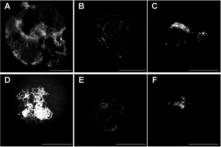
Figure 4. Immunofluorescent images of abnormal oocytes in fully-grown germinal vesicle-stage oocytes. Shown are single confocal sections of germinal vesicle (GV) or oocytes. A. Misshapen and torn oocyte from aged female stained for DNMT1. B-D. GV of oocytes from aged mice displaying a distorted nuclear structure stained for H3K9me2 (B and C) and DNA (D). E-F. GV of oocytes displaying DNA circles from a young mouse and aged mouse stained for H3K9me2 and H4K5ac, respectively. Scale bar = 50 µm.

