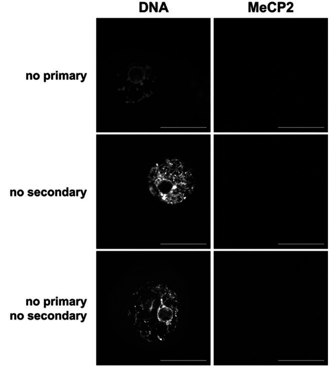
Supplemental Figure 1. Immunofluorescent no antibody control images for MeCP2 staining. Shown are single confocal sections of the nucleus (GV) without exposure to primary antibody (top ), secondary antibody (middle), or either antibody (bottom ). Confocal settings were the same within each technical replicate. DNA stain = YoYo1 Iodide. Scale bar = 50 µm.

