. 2021 Jun 2;10:e66739. doi: 10.7554/eLife.66739

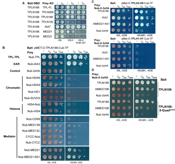
Figure 2. The Helix 8 repression domain of TPL directly interacts with AtMED21 and AtMED10B.

(A–C, E) Cytoplasmic split-ubiquitin system (CytoSUS) assays with candidate interacting proteins. Nub-3xHA is the N-terminal fragment of ubiquitin expressed with no fusion protein and is used as a negative control. Each prey protein is from Arabidopsis. -WL, -ADE: dropout lacking Trp, Leu, and Ade (growth control); -WLMH, -ADE: dropout lacking Trp, Leu, His, Met, and Ade (selective media). The plating for each panel was performed at the same day, white lines are provided when plates were cropped for clarity. (B) Alignments of the Arabidopsis (At) and Saccharomyces (Sc) MED21 proteins are shown above cytoSUS assays with the same bait shown in (A). Western blots below the colonies indicated that AtMED21 N-terminal Δ3 and Δ5 are well expressed in assay conditions. (C) CytoSUS assays with selected Mediator proteins in the middle module. (D) The TPL-ProteinA-TF fusion protein can pull down TPL, AtMED21, and AtMED10B from yeast extracts using IgG-beads. Detection of the VP16 transcriptional activator demonstrates enrichment of the fusion protein (αVP16). Each prey protein is detected via the 3xHA tag (αHA), and efficacy of purification was judged by PGK1 depletion (αPGK1). (E) A TPL-N truncation lacking the LisH domain (TPLH2-H9) could still interact with the AtMED21-N31 truncation. This bait construct interacted with IAA3, but only minimally with the negative control (free Nub-3xHA). (F) Yeast Mediator (bottom, 5N9J) and AtTPL (top, 5NQV) manually juxtaposed to compare relative domain sizes and feasibility of a TPL-MED21-MED10B interaction. TPL Helix 8–9 is colored green. MED21 is colored aqua, with the N-terminus colored red, and the IAA27 EAR peptide in orange. MED10 is colored teal, with the C-terminus colored purple. The dotted line indicates the border between TPL and Mediator structures.
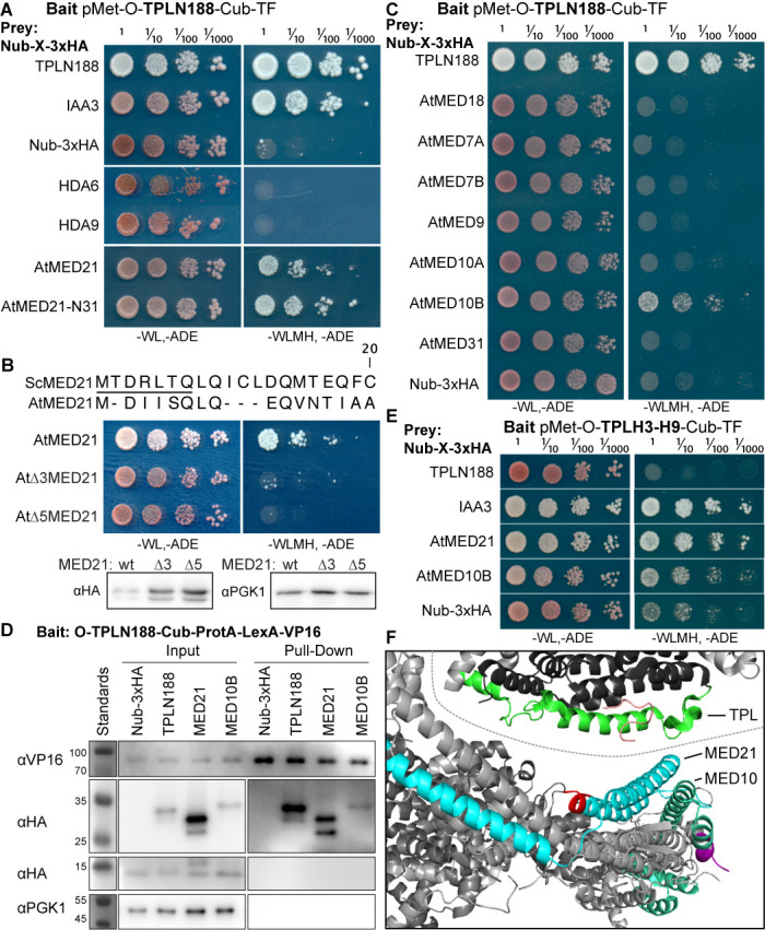
Figure 2.

Figure 2—figure supplement 1. The TPL-N terminal domain (TPLN188) interacts with the N-terminus of AtMED21.

Figure 2—figure supplement 1.

(A) Identifying TPL-N terminal domain interactor proteins through yeast two hybrid screening identifies TPL as a problematic bait protein as it may silence the activation despite successful binding of a prey protein (see second row from bottom where N188 and MED21 show very weak reporter activity on 3AT). 3AT: 3-amino-1,2,4-triazole. Plates were measured after 3 days to allow TPL-MED21 interactions to grow. (B) Identifying TPL-N terminal domain interactor proteins through cytoplasmic split ubiquitin protein interaction assay. We tested the N-terminal and C-terminal portions of MED13 separately and divided the coding sequence at amino acid 967 (MED13N = aa1-967, MED13C = aa968-1908). Each bait tested is the Arabidopsis homolog cloned from cDNA from the Col-0 accession, with the exception of AtMED13, which was synthesized de novo via Twist (https://www.twistbioscience.com/). Plates were scanned at 3 days after plating to allow weaker interactions to develop if they were present. (C) TPL interacts with MED21 through an interaction within Helices 8–9. Plates were scanned at 2 days after plating. (D) The Helix 8 Quadruple mutation (V145A, E146A, K148A, K149A) does not affect AtMED10B binding to TPL. Plates were scanned at 3 days after plating. (B–D) The relative position of the N-terminal portion of ubiquitin (Nub) is indicated for each bait protein.

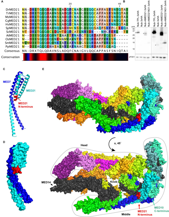
Figure 2—figure supplement 2. Homology and structure of the MED21 subunit of the Mediator complex.

Figure 2—figure supplement 2.

(A) Protein alignment of selected MED21 homologs from various species. Dr: Drosophila melanogaster; Tr: Takifugu rubripes; Ms: Mus musculus; Gg: Gallus gallus; Hs: Homo sapiens; Sp: Strongylocentrotus purpuratus; Sc: Saccharomyces cerevisiae; At: Arabidopsis thaliana; Os: Oryza sativa; Sm: Selaginella moellendorffii; Pp: Physcomitrella patens. Alignment was performed in CLC sequence viewer 7 using a neighbor joining method. (B) Protein levels of AtMED21 cytoplasmic split-ubiquitin system (cytoSUS) constructs in yeast. Two different exposure times are shown to demonstrate the lower abundance of the truncation with only the first 31 amino acids of the AtMED21 (N31). Asterisks indicate the size predicted for the indicated protein. (C, D) Structure of the MED21 (cyan) and MED7 (blue) hetero dimer, adapted from 1YKH (Baumli et al., 2005). The amino acids in the N-terminus that were solved are highlighted in red up to the 7th amino acid of the yeast MED21. (C) The cartoon visualization (D) Surface visualization. (E) Core mediator (5N9J, Nozawa et al., 2017) with the location of MED21 and MED7 indicated with the same colors from (C, D). In this structure, the location of the MED21 N-terminus is again indicated in red, demonstrating its close proximity to the Knob region (dotted circle).