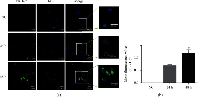
Figure 3.

PKH67-labeled MI-associated EVs (green) taken up by HUVECs after coculture for 24 h and 48 h. (a) Immunofluorescence images were obtained using a laser scanning confocal microscope (at the magnification of 400x). (b) Mean fluorescence values of PKH67 analyzed by ImageJ software. ∗P < 0.05, compared with cultured for 24 h, n = 3.
