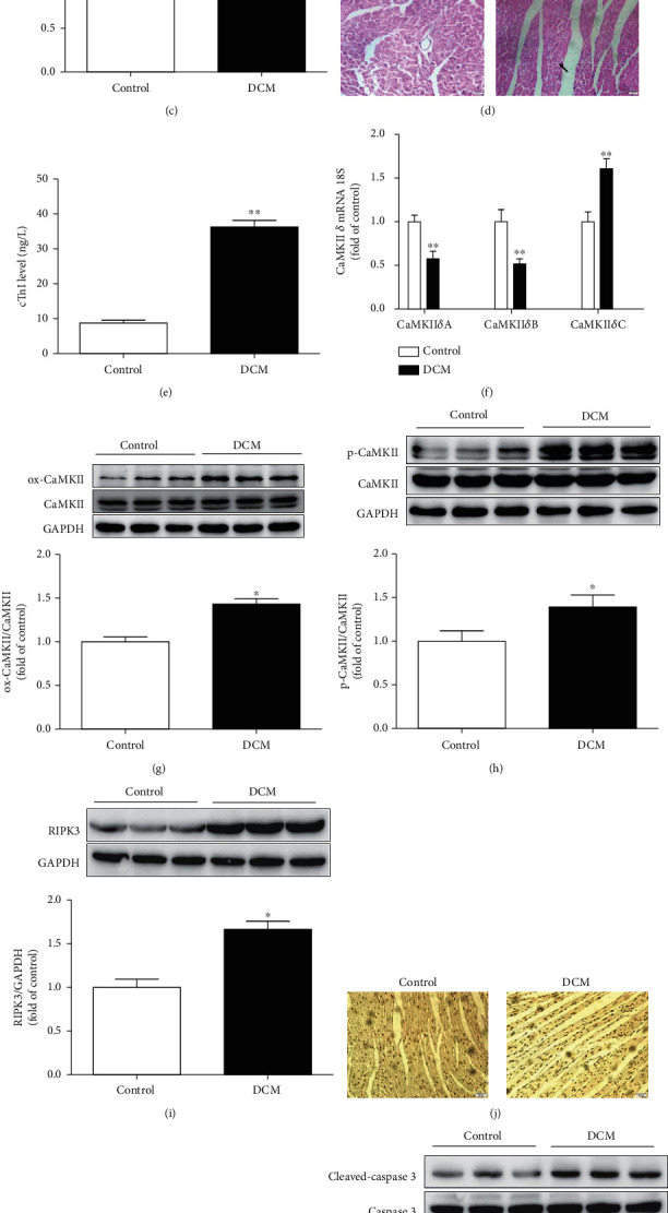
Figure 1.

Cardiac dysfunction, CaMKIIδ activity, and necroptosis are augmented in DCM. Male C57BL/6 mice were injected with 60 mg/kg/d STZ for 5 consecutive days after a 12-hour overnight fast. Mice in the control group were injected with the same amount of citrate buffer. (a–c) Cardiac function was assessed by echocardiography, and EF, FS, and E/A were calculated. (d) Myocardium injury was measured by HE staining. Bar = 20 μm. (e) cTnI was detected. (f) The mRNA levels of CaMKIIδA, CaMKIIδB, and CaMKIIδC of the myocardium were detected by quantitative real-time PCR. 18S was serviced as a housekeeping mRNA. (g–h) Expression of CaMKII oxidation (ox-CaMKII), CaMKII phosphorylation (p-CaMKII), and total CaMKII was quantified by western blot. GAPDH was used as a loading control. (i) RIPK3 protein expression was quantified by western blot. GAPDH was used as a loading control. (j, k) Cell apoptosis of myocardium was detected with TUNEL staining and quantified with Image J analysis software. Bar = 50 μm. (l) Cleaved-caspase 3 and caspase 3 protein expression were quantified by western blot. GAPDH was used as a loading control. ∗P < 0.05 and ∗∗P < 0.01, significantly from control, n = 6.
