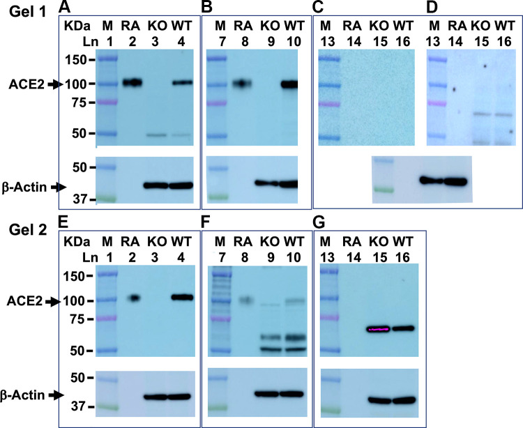
Figure 5.
Commercial angiotensin-converting enzyme 2 (ACE2) antibody sensitivity and specificity. Shown are representative Western blots from two gels with 16 wells each. After protein transfer, the PVDF membranes contained protein markers (M; lanes 1, 6, and 13), mouse recombinant ACE2 protein (RA; lanes 2, 8, and 14), and renal tissue from ACE2 knock out (KO; lanes 3, 9, and 15) and wild-type (WT; lanes 4, 10, and 16) female C57BL/6 mice. Membranes were cut into three pieces each and blotted with either anti-mouse ACE2 antibodies from: R&D (A), Abcam (B), and Santa Cruz (C) or anti-human ACE2 antibodies from: Cell Signaling (E), Novus (F), and, Proteintech (G). Super Signal West Pico Plus substrate was used to detect ACE2 immunoreactive protein in A–C and E–G. D shows membrane exposed to the Santa Cruz antibody (C) re-probed with the Femto maximum sensitivity substrate. After probing with ACE2 antibodies, all membranes were stripped and incubated with antibodies to β-actin to control for differences in protein loading.

