Fig. 1.
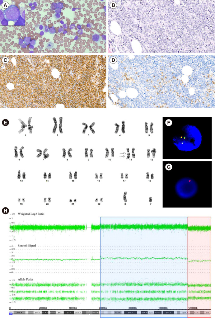
Bone marrow findings in Burkitt-like lymphoma with 11q aberration. (A) Bone marrow aspirate smear revealed medium- to large-sized lymphoma cells with finely clumped chromatin, variably prominent nucleoli, moderate amounts of deeply basophilic cytoplasm, with or without lipid vacuoles (Wright-Giemsa stain, ×400 and ×1,000). (B) Bone marrow biopsy section revealed diffuse infiltration of lymphoma cells composed of medium- to large-sized cells (H & E stain, ×200). On immunohistochemical staining, (C) lymphoma cells were diffusely positive for CD20, and (D) reactive T cells were positive for CD3 (×200). (E) Chromosome analysis showed an abnormal chromosome 11, including inverted duplication of the part of the long arm between 11q24 and 11q13 and terminal deletion of the long arm from 11q24 to 11ter (arrow). On interphase fluorescence in-situ hybridization, (F) KMT2A (MLL) Dual-Color Break Apart probe (Vysis) showed three green/orange (yellow) fusion signals indicating a gain of 11q23.3, and (G) Telomere 11q SpectrumOrange probe (Vysis) showed a single orange signal, indicating a loss of 11q24. (H) Chromosomal microarray analysis showed a copy-number gain of 11q12.2 q23.3 (blue) followed by an adjacent distal loss of 11q23.3 q25 (red).

