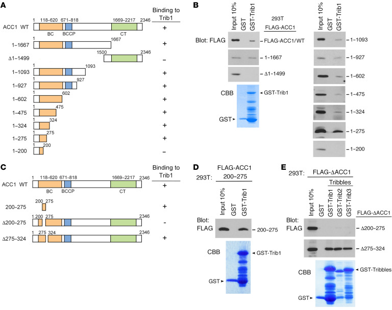
Figure 2. Identification of the Tribbles-binding site in ACC1.
(A) Schematic representation of ACC1 deletion mutants. The results of Trib1 binding are summarized on the right. (B) GST-control and GST-Trib1 fusion proteins were incubated with 293T cell lysates containing FLAG-tagged ACC1WT and deletion mutant proteins. Bound proteins were detected by immunoblotting with an antibody against a FLAG epitope. The GST-Trib1–fused protein was visualized by CBB staining to evaluate its amount. (C) Schematic representation of ACC1 deletion mutants minimized for ACC1-Trib1 binding. The results of Trib1 binding are summarized on the right. (D and E) GST-control and GST-Trib1 fusion proteins were incubated with 293T cell lysates containing FLAG-ACC1/200–275 (D). GST-control and all GST-Tribbles (GST-Trib1, GST-Trib2, and GST-Trib3) fusion proteins were incubated with 293T cell lysates containing FLAG-ACC1/Δ200–275 and ACC1/Δ275–324 (E). Bound proteins were detected by immunoblotting with an antibody against a FLAG epitope. GST-Tribbles–fused proteins were visualized by CBB staining to evaluate their amounts.

