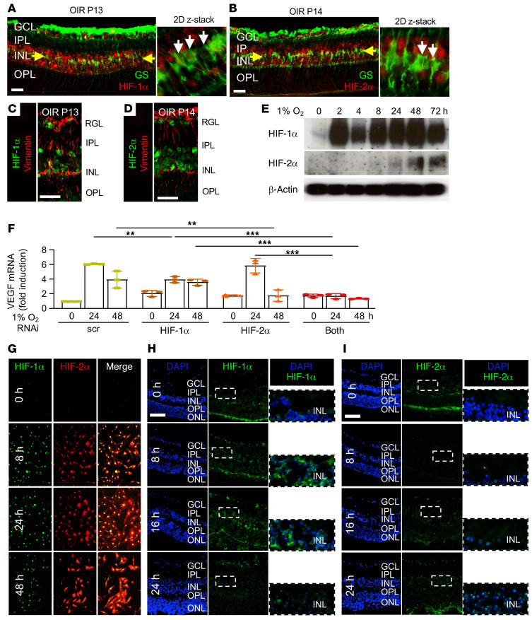
Figure 10. Expression of HIF-1α and HIF-2α in OIR mice Müller cells and mouse retinal explants treated with hypoxia.
(A–D) IF demonstrating the expression of HIF-1α (A and C) and HIF-2α (B and D) in Müller cells expressing glutamine synthetase (GS; A and B) or vimentin (C and D) in the INL (yellow arrows) at P13 or P14. White arrows point to cells coexpressing GS and HIF-1α or HIF-2α. (E) Expression of HIF-1α and HIF-2α over time by Western blot in primary mouse Müller cells treated with hypoxia. (F) Expression of VEGF mRNA expression in hypoxic primary mouse Müller cells after RNAi knockdown of HIF-1α, HIF-2α, or both. (G) Coexpression of HIF-1α and HIF-2α by IF in primary mouse Müller cells treated with hypoxia. (H and I) Rapid but transient expression of HIF-1α (H) and delayed expression of HIF-2α (I) in adult mouse retinal explants treated with hypoxia for 8 to 24 hours. Data are shown as mean ± SD. Statistical analyses were performed by 2-way ANOVA with Bonferroni’s multiple-comparison test. *P < 0.05; **P < 0.01; ***P < 0.001. n = 4 to 6 animals. GCL, ganglion cell layer; IPL, inner plexiform layer; INL, inner nuclear layer; OPL, outer plexiform layer; ONL, outer nuclear layer; h, hours. Scale bar: 100 μm.

