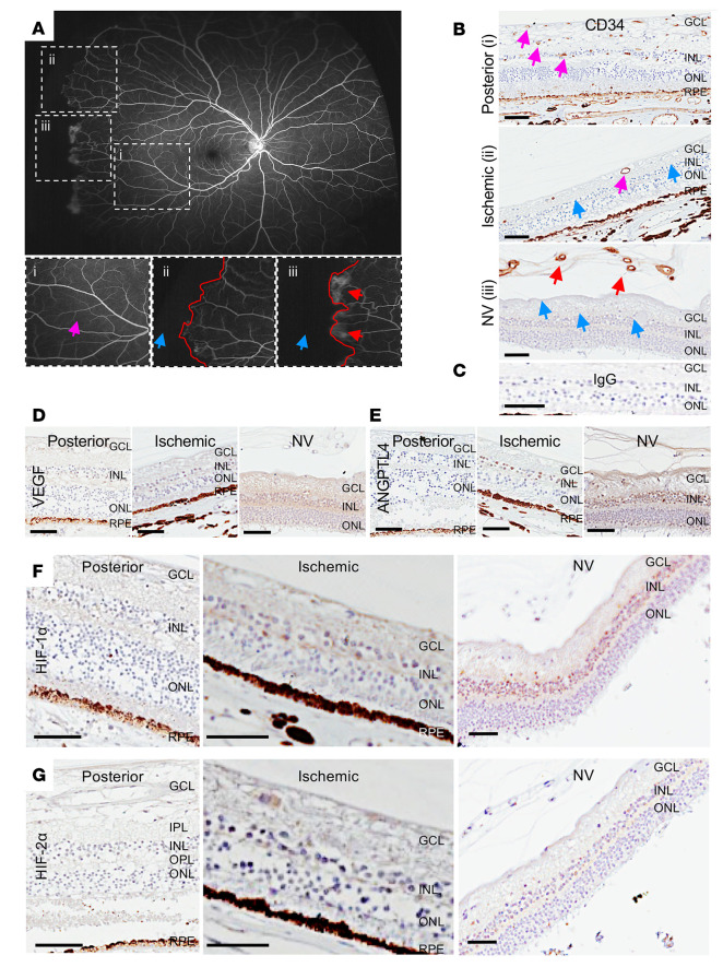
Figure 2. Expression of HIF-1α and HIF-2α and HIF-regulated angiogenic mediators in the ischemic retina of patients with sickle cell disease.
(A) Fluorescein angiogram from a patient with PSR demonstrating areas of normal perfusion (pink arrow) posteriorly (i) and nonperfusion (blue arrow) peripherally (ii and iii); the margin between perfused and nonperfused retina demarcated in red. Areas without (ii) and with (iii) retinal NV (red arrows) are seen adjacent to one another. (B) Representative images from immunohistochemical analysis for the endothelial cell marker CD34 in posterior perfused and peripheral ischemic retina, the latter without and with NV (red arrows). In the posterior retina, vessels are noted in the superficial, intermediate, and deep vascular plexuses (pink arrows). In the ischemic retina (without NV), rare vessels are noted in the superficial and intermediate vascular plexuses (pink arrows), but vessels are absent in the deep vascular plexus (blue arrows). In ischemic retina with overlying retinal NV (red arrows), there was a complete absence of retinal vessels in all vascular plexuses (blue arrows). (C) IgG was used as a negative control. (D–G) Expression of the HIF-regulated angiogenic mediators VEGF (D) and ANGPTL4 (E), HIF-1 (F), and HIF-2α (G) in these same regions of sickle cell eyes. n = 5 eyes. GCL, ganglion cell layer; IPL, inner plexiform layer; INL, inner nuclear layer; OPL, outer plexiform layer; ONL, outer nuclear layer; RPE, retinal pigment epithelium. Scale bar: 100 μm.

