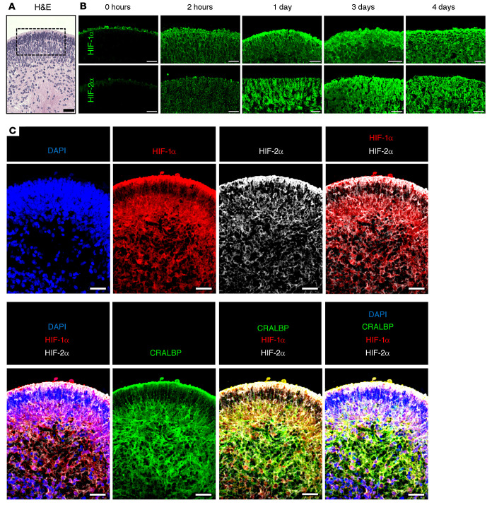
Figure 4. Simultaneous expression of HIF-1α and HIF-2α in hypoxic retinal cells in hiPSC-derived 3D retinal organoids.
(A) H&E staining of D120 3D retinal organoid prior to treatment with prolonged hypoxia. (B) Expression of HIF-1 and HIF-2α over time in boxed region (from A) of D120 3D retinal organoids exposed to hypoxia for 2 hours to 4 days. (C) HIF-1α, HIF-2α, and CRALBP expression in hypoxic hiPSC-derived retinal organoids treated with hypoxia. Scale bars: 25 μm.

