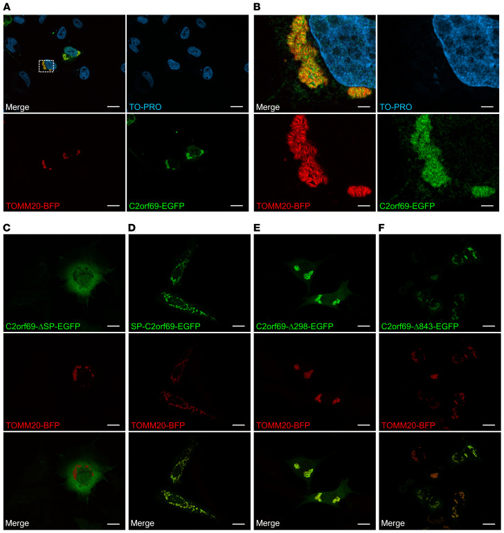
Figure 3. C2orf69 shows mitochondrial localization.
COS-7 cells were transfected with the indicated constructs, and nuclei were stained with TO-PRO. Localization of C2orf69‑EGFP (A), C2orf69‑ΔSP‑EGFP (C), SP‑C2orf69‑EGFP (D), C2orf69‑Δ298‑EGFP (E), C2orf69‑Δ843‑EGFP (F), and the mitochondrial marker TOMM20‑BFP. Scale bars: 20 μm. (B) Higher-magnification images of the mitochondria from A. Scale bars: 3.5 μm. Representative images are shown (n = 3).

