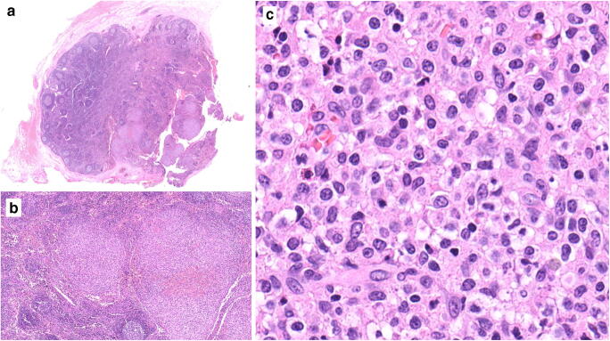
Fig. 1.
Histopathology of the lymph node. a Panoramic view showing partial replacement of the lymphoid tissue by relatively circumscribed nodules (lower right). Elsewhere, the nodal architecture is preserved with follicular hyperplasia. b The nodules are composed of monotonous and medium-sized lymphoid cells. c Cytological features at high power. Hematoxylin and eosin stain; original magnifications × 8, × 50, and × 400, respectively

