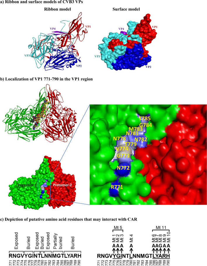
Figure 1.
Structural characteristics of VP1 771–790 of CVB3 showing their relationship to the binding region between viral canyon and CAR, and an approach to create mutant viruses. (a) Ribbon and surface models of CVB3 VPs. Using the published pentamer structure of CVB3 (PDB: 1COV)16,17, all four structural proteins of CVB3 (VP1 [red], VP2 [blue], VP3 [turquoise], and VP4 [purple]) were localized in the protomer, as illustrated in the ribbon (left panel) and surface (right panel) models. (b) Localization of VP1 771–790 in the VP1 region. The amino acid residues of VP1 771–790, depicted in balls between two protomers (green and red) are shown in the top panel, and their positions are indicated in yellow. The surface model (bottom panel) shows the exposed amino acid residues that may potentially interact with the CAR; their positions are shown in yellow in the inset. The interactions of VP1 771–790 with CAR were analyzed using the BIOVIA Discovery Studio v4.0 (https://www.discngine.com/discovery-studio). (c) Depiction of putative amino acid residues that may interact with CAR. After localizing the residues within the VP1 region in relation to CAR as described above, the putative residues that potentially interact with the host receptor CAR were identified as exposed residues (RNGV, NT, and NN), whereas YGI, L, and LYARH were identified as buried residues, which are not expected to interact with CAR (left panel). The right panel represents mutations introduced at various positions individually or in combination.

