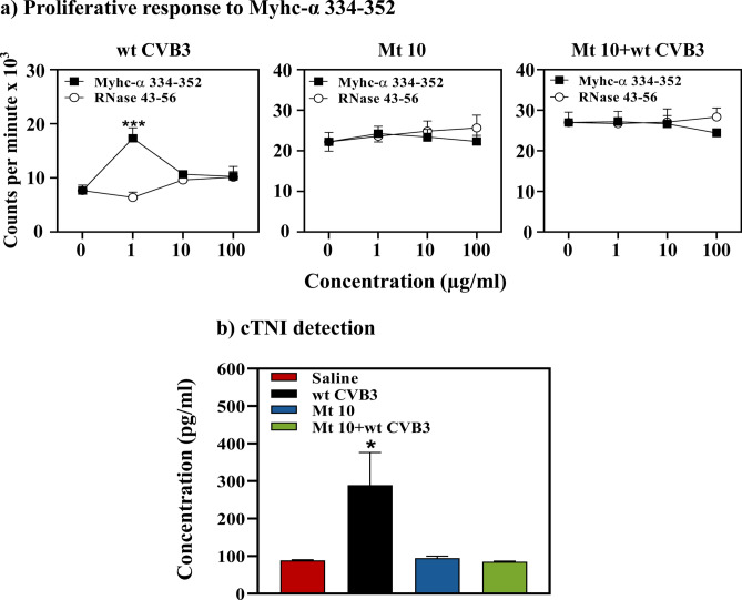
Figure 8.
Mice injected with Mt 10 virus did not reveal autoimmunity or cardiac injury. (a) Proliferative response to Myhc-α 334–352. Groups of mice were administered saline or Mt 10 virus, and after 14 days, they were challenged with or without CVB3. Three weeks later, lymphocytes were prepared, and cells were stimulated with or without Myhc-α 334–352 or control (RNase 43–56) for 2 days. After pulsing with 3[H] thymidine for 16 h, proliferative responses were measured as cpm. Mean ± SEM values obtained from three individual experiments, each involving n = 3 to 8 mice, are shown. (b) cTnI detection. Undiluted sera obtained from the above groups were added in duplicates to pre-coated, cTnI ELISA strip plates. After incubation and a series of washes, HRP-conjugated rabbit anti-mouse antibody was added followed by TMB substrate and stop solution. Plates were read at 450 nm to obtain OD values, and concentrations of unknown samples were determined using the standard curve. Mean ± SEM values representing 6 samples per group, each containing n = 3 to 5 mice, are shown. Unpaired Student’s t-test (two-tailed) was used to determine significance between groups [for panels (a,b)]. *p ≤ 0.05, and ***p ≤ 0.001.

