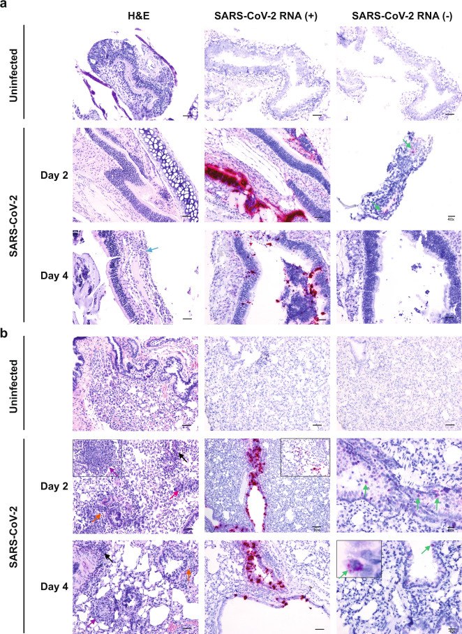
Fig. 3. Histopathology and virus distribution.
Hematoxylin/eosin (H&E) staining (left) and in situ hybridization (ISH) using antisense probes that detect the SARS-CoV-2 genome/mRNA (middle) and sense probes that detect anti-genomic RNA (right) were carried out on (a) nasal turbinates and (b) lung tissue of uninfected and SARS-CoV-2-infected deer mice (105 TCID50 i.n. route) at 2 and 4 dpi. Positive detection of viral genomic RNA/mRNA or anti-genomic RNA is indicated by magenta staining (middle/right panels and insets). Arrows indicate perivascular infiltrations of histiocytes and neutrophils (black), peribronchiolar infiltrations of histiocytes and neutrophils (orange), mild neutrophilic infiltration in the submucosa (blue), occasionally observed discrete foci of interstitial pneumonia (purple), occasionally observed multinucleated syncytial cells (magenta), and anti-genomic RNA occasionally in individual scattered bronchiolar epithelial cells (green and right inset). The magnification is ×20 for H&E and ×20 for ISH unless otherwise indicated. Scale bars =100 μm, 50 μm, and 20 μm for 10×, 20×, and 40×, respectively. A total of three biological replicates (individual animals) were assessed at each time point, and the selected panels are representative of these findings.

