Figure 4.
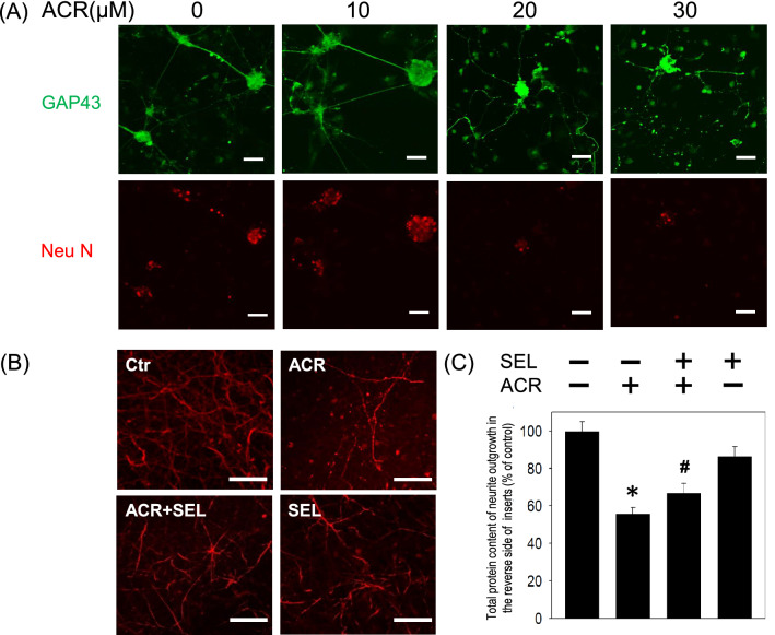
Effect of selumetinib on acrolein-induced reduction in neurite outgrowth. (A) Primary cultured cortical neurons (3 × 10⁵ cells in a 35 mm petri dish containing coverslips) were treated with acrolein (0–30 μM) for 24 h. Representative immunofluorescent data using GAP-43 antibody show the effect of acrolein on neurite outgrowth. Caliber: 50 μm. (B) Primary cultured cortical neurons were cultured in Transwell inserts and treated with acrolein (30 μM) with/without selumetinib (SEL, 10 μM) for 24 h. Representative immunofluorescent data using MAP-2 antibody show the neurites on the reverse side of the Transwell insert. Caliber: 50 μm. (C) Statistical data showed the effect of selumetinib on acrolein-induced reduction in total protein content of neurite outgrowth grown on the other side of Transwell inserts. Values are the mean ± SEM. (n = 3/treatment). *p < 0.05 statistically significant in the acrolein groups compared with the control groups; #p < 0.05 statistically significant in acrolein plus SEL group compared with acrolein group by one-way analysis of variance (one-way ANOVA) and followed by the LSD test as post-hoc method.

