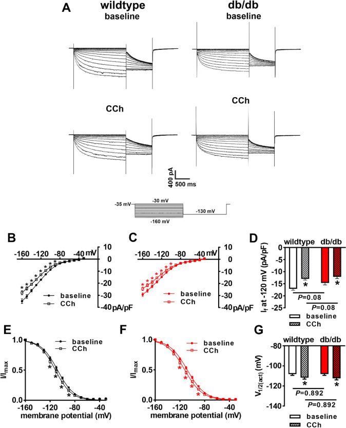
Figure 5.
Effects of carbachol on the hyperpolarization-activated current (If) in db/db SAN myocytes. (A) Representative If recordings at baseline and after CCh (10 µM) application in wildtype and db/db SAN myocytes. Voltage clamp protocol shown at bottom of recordings. (B) If IV curves at baseline and after CCh application in wildtype SAN myocytes (n = 15). (C) If IV curves at baseline and after CCh application in db/db SAN myocytes (n = 16). For panels (B) and (C) *P < 0.05 vs. baseline by two-way repeated measures ANOVA with Holm–Sidak posthoc test. (D) If density at − 120 mV at baseline and after CCh application in wildtype and db/db SAN myocytes. *P < 0.05 vs baseline by two-way ANOVA with Holm–Sidak posthoc test. (E) If steady-state activation curves at baseline and after CCh application in wildtype SAN myocytes (n = 15). (F) If steady-state activation curves at baseline and after CCh application in db/db SAN myocytes (n = 16). (G) If V1/2(act) at baseline and after CCh application in wildtype and db/db SAN myocytes. *P < 0.05 vs. baseline by two-way ANOVA with Holm–Sidak posthoc test. Refer to Supplementary Table 1 for additional analysis of If steady-state activation.

