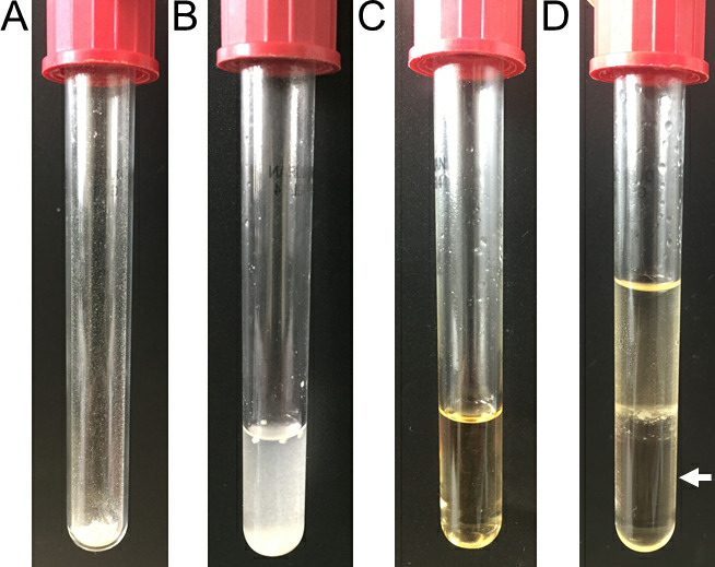
Figure 6. Images of the acidic methanolysis steps of PHB.

Weighted cells (A), complete methanolysis sample before oil bath (B), sample after oil bath (C) and phase-separated sample with the bottom organic phase (white arrow, D).

Weighted cells (A), complete methanolysis sample before oil bath (B), sample after oil bath (C) and phase-separated sample with the bottom organic phase (white arrow, D).