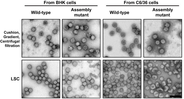
Figure 4. Purification of wild-type and assembly mutant virus.
Sindbis virions (wild-type and an assembly mutant) from both BHK-infected and C6/36-infected cells were purified using two protocols and then imaged by TEM. The top row shows the virion purification using three sequential steps: pelleting through a sucrose cushion, applying the pellet on a sucrose gradient, and buffer exchange and concentration of the viral band using centrifugal filtration. The bottom row shows the virus samples from the one step virion purification using the LSC protocol. In addition to having more particles on the grid, the LSC purified particles show fragile particles as indicated by the dent in the middle of the particle (arrow). These particles are most likely degraded or further damaged during the harsher purification methods. As a result, the diversity of assembly mutants is not represented in the upper panels. Scale bar (shown bottom right image) is 200 nm and is applicable to all EM images.

