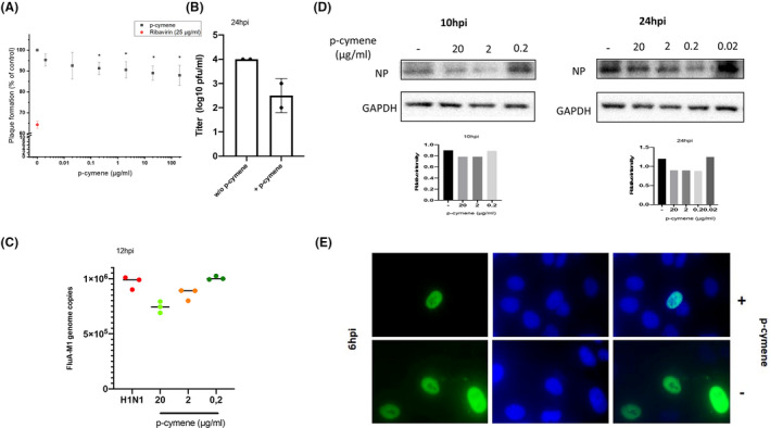
FIGURE 5.

(A) Plaque reduction assay of Madin‐Darby Canine Kidney (MDCK) cells infected with Influenza H1N1 virus (0.05 PFU/cell) and incubated with variable concentrations of p‐cymene for 72 h. Figure shows mean ± SD of two experiments in triplicates. Ribavirin (red dot) at 25 μg/ml is presented as a positive control. *p < .05 as compared to non‐treated cells. (B) p‐cymene reduces the production of progeny virus, as assayed by quantitative PCR of the supernatants of MDCK cells were pretreated with p‐cymene (20 μg/ml) and infected with influenza A/H1N1 virus (0.1 PFU per cell) for 24 h. (C) Quantification by qRT‐PCR of FluA M1 genome copies in MDCK cells infected with FluA/H1N1 at MOI 0.5 PFU/cell. Data shown are means ± SD of two independent experiments. (D) Immunoblot analysis of influenza nucleoprotein (NP) in total MDCK cell lysates incubated for 10 and 24 h post‐infection with variable concentrations of p‐cymene. (E) The role of p‐cymene in the NP distribution. MDCK cells were infected with FluA/H1N1 (MOI 5) in the absence or presence of p‐cymene. Six hours after the infection, cells were fixed and subsequently stained using influenza A anti‐NP primary antibody followed by FITC secondary antibody (green). DAPI was used to visualize the nucleus area (blue)
