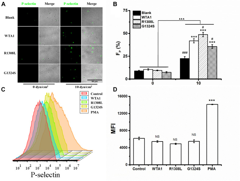
FIGURE 2.
P-selectin translocation of platelets adhered to either immobilized- and suspended-A1 domain of von Willebrand factor and its two mutants (R1308L and G1324S) with or without mechanical stimuli for 8 min. (A) P-selectin immunolocalization (green) and (B) P-selectin-positive fraction (FP) of platelets on substrates coated with WTA1, R1308L, and G1324S without or with fluid shear stress stimulus of 10 dyn/cm2 for a stimulus time of 8 min. Merged images of differential interference contrast and green fluorescence are shown with bar = 50 μm. The data represent the mean ± SEM from three independent experiments. Statistical significance was analyzed by two-way ANOVA for multiple comparisons with Bonferroni post hoc test; ***p < 0.001 compared with the blank group, #p < 0.05 and ###p < 0.001 compared with WTA1 group, and ΔΔΔp < 0.001 compared with 0 dyn/cm2 group. (C) Representative flow cytometry histograms of P-selectin translocation for platelets treated without (control) or with WTA1, R1308L, G1423S, and PMA. (D) Bar graph representing the mean fluorescence intensity of P-selectin-positive platelets (MFI) in various treatments from flow cytometry. All data are shown as mean ± SEM from three independent experiments and analyzed by one-way ANOVA for multiple comparisons. ***p < 0.001; NS, not significant compared with the control group.

