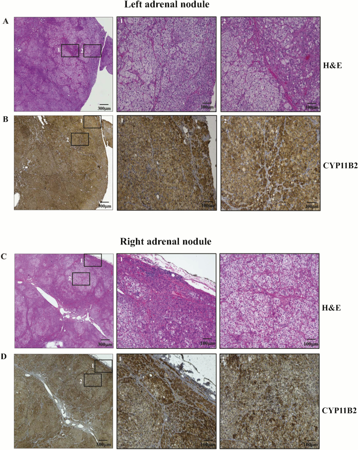
Figure 3.
Representative histopathological analysis of two adrenal nodules of a KCNJ5 mosaic individual. Hematoxylin-eosin staining of a left adrenal nodule (a). Immunostaining for CYP11B2 of a left adrenal nodule (b). Hematoxylin-eosin staining of a right adrenal nodule (c). Immunostaining for CYP11B2 of a right adrenal nodule (d). Corresponding amplified images from the squares numbered with 1 and 2 in the lower magnification field can be seen in the adjacent images numbered as 1 and 2. Slides were scanned in a ×10 objective.

