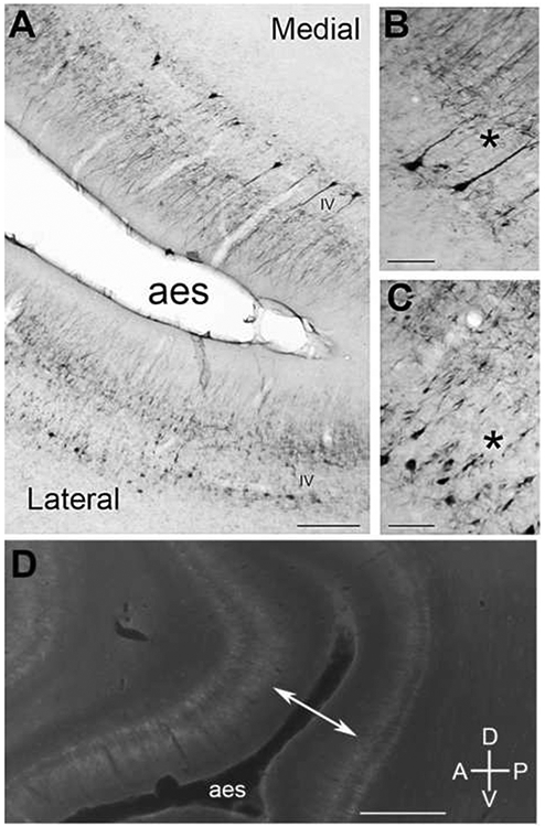
Figure 13.

SMI-32 expression in the anterior ectosylvian (AE) sulcus. A: The photomicrograph shows possible SMI-32 subregions within the AE sulcus, medial and lateral. B: At higher power, a characteristic of the medial portion of the AE sulcus is the more full and reactive apical dendrites (*). C: The lateral portion of the AE sulcus has a more fragmented SMI-32 reactivity throughout layer V somata. D: Expression profile of the AE sulcus from another brain prepared sagittally. The double-headed arrow points to reactive disparity evident between supragranular layers of the anterior and posterior regions of the AE sulcus. Scale bars: A = 1000μm; B&C = 50μm; D = 2000μm.
