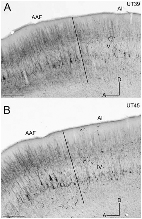
Figure 5.

SMI-32 immunoreactivity in sagittal sections through the middle ectosylvian gyrus. Layer IV is labeled in each and borders are marked by black lines. A: The AI/AAF border at x200 in case UT39. B: The AI/AAF border at x200 in case UT45. D=dorsal; A=anterior. Scale bars: A&B = 1000μm.
