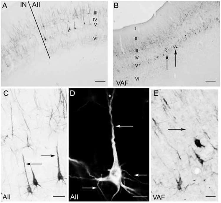
Figure 8.

SMI-32 reactivity in the middle ectosylvian areas AII (A,C,D) and VAF (B,E) A: Border of AII and neighboring cortical region IN. B: Low magnification photomicrograph of SMI-32 expression in VAF. Note heavy reactivity in layer V somata (arrows). C: Magnification (x100) of AII layer V somata. Arrows indicate the characteristic well labeled apical dendrites. D: A layer V pyramidal cell in AII at x600 under darkfield illumination. Arrows indicate the well defined dendrites that were commonly identified. The asterisk indicates a commonly observed bifurcation in an apical dendrite. E: Layer V somata in VAF at x100. The arrow shows the lack of a well-labeled apical dendrite, which is commonly absent from layer V somata in VAF. Scale bars: A and B=500μm; C and E=50μm; D=40μm.
