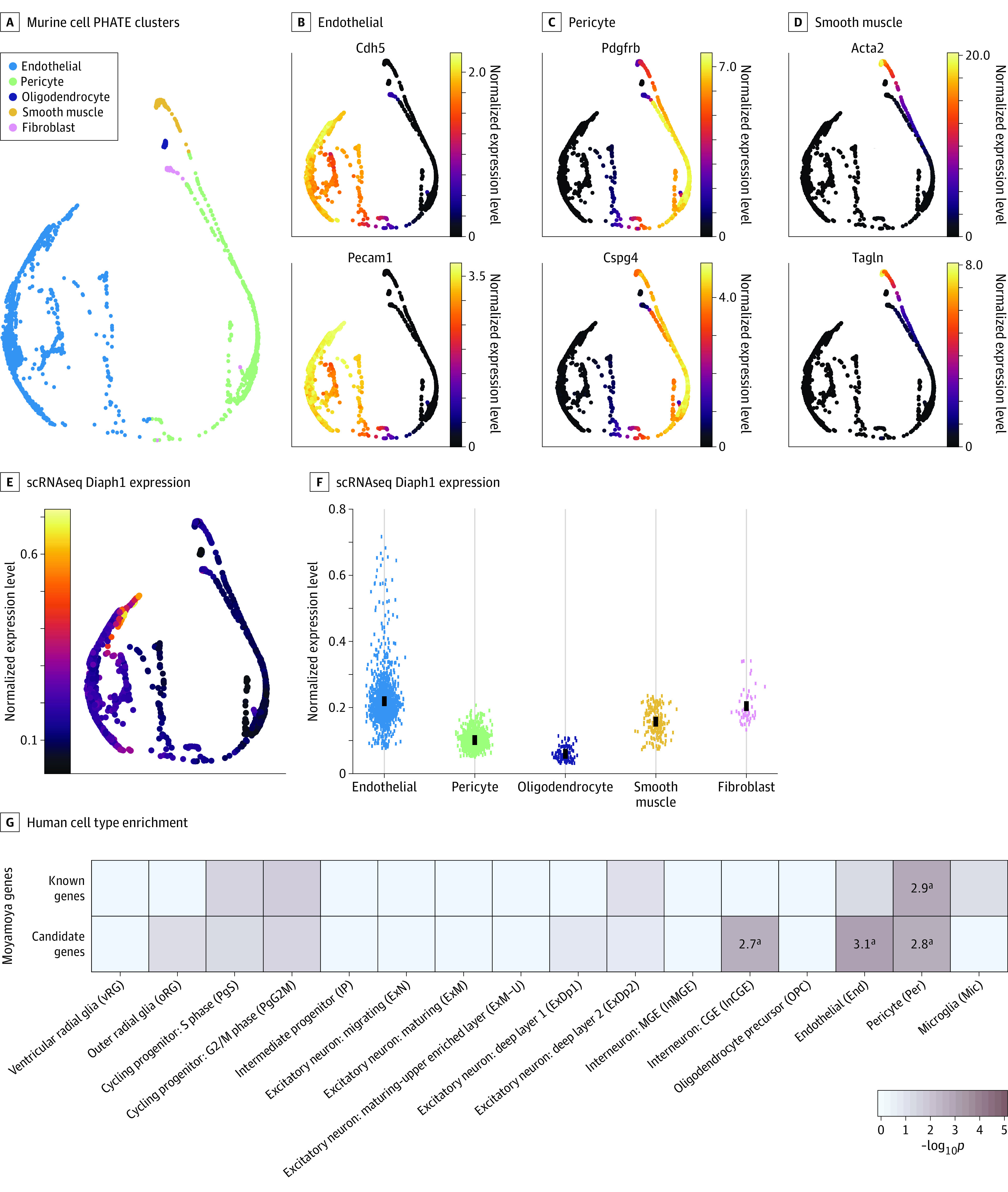
Figure 2. Enriched Expression of DIAPH1 and Other Moyamoya Disease Risk Genes in Vascular Cells.

A-D, Cell type–specific clusters in mouse brain vascular cells using Betsholtz scRNaseq data. A, Potential of heat-diffusion for affinity-based transition embedding (PHATE) plot clusters of endothelial cells, pericytes, oligodendrocytes, smooth muscle cells, and fibroblasts. B-D, PHATE plots of 2 marker genes per cluster. B, Endothelial cells expressing Cdh5 and Pecam1. C Pericytes expressing Pdgfrb and Cspg4. D, Smooth muscle cells expressing Acta2 and Tagln. E, PHATE plot of cell type–specific expression of Diaph1. Expression maps best to endothelial cells. F, Diaph1 messenger RNA abundance in each cell cluster. G, Enrichment analysis across cell type markers of the midgestational human brain 69 for known human genes and cohort-determined candidate risk genes for moyamoya disease.
aRepresents significant enrichment at the Bonferroni multiple-testing cutoff (α = .05/16 = 3.13 × 10⁻³).
