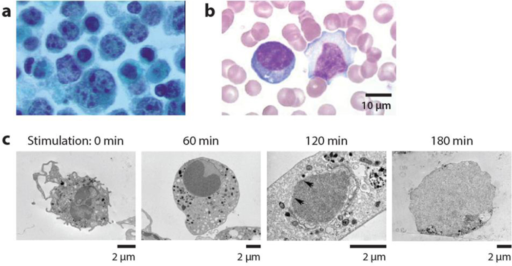
Figure 2.
Changes occurring in cellular structure with disease. (a) Lymphoma cells present with abnormally shaped nuclei, with overall light staining of chromatin and dense staining of punctate chromatin. Figure adapted from Reference 60 with permission. (b) Reactive lymphocytes with expanded basophilic cytoplasm and irregular nuclear shape. In contrast, resting lymphocytes have reduced cytoplasm and regular nuclear contours (http:www.wikidoc.orgindex.phpReactive_lymphocyte). (c) Neutrophils undergoing a process of NETosis, a form of programmed cell death in which the nuclear membrane disassembles and chromatin is released. The nuclear envelope dissolves and chromatin mixes with granules during a 3-hour period (67).

