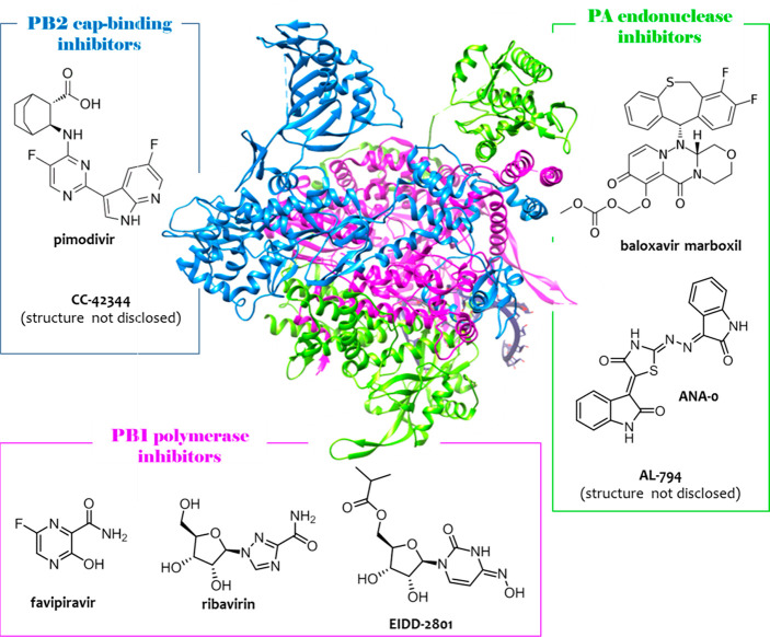
Figure 1.
Crystal structure of flu A RdRP determined from bat flu A H17N10 strain (pdb: 4WSB(30)) and chemical structures of inhibitors of PA (green), PB2 (blue), and PB1 (magenta) subunits, approved or in the pipeline. The overall RdRP is U shaped with the PAN endonuclease and PB2 cap-binding domains being the two upper protuberances, the PAC domain being the bottom, and the PB1 polymerase domain filling the interior. Among the reported compounds, PA endonuclease inhibitor baloxavir marboxil and PB1 inhibitor favipiravir have been approved. The figure is author created, and the RdRP structure has been adapted from the pdb mentioned above and drawn by using UCSF Chimera package.47

