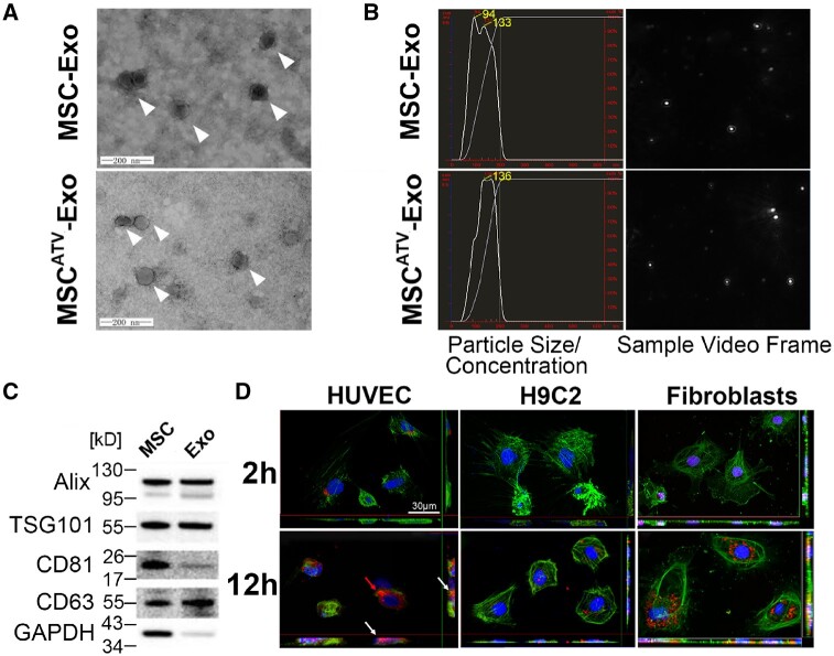
Figure 1.
Characterization and functional validation of exosomes derived from ATV-treated MSCs. (A) Cup-shaped morphology of purified MSC-Exo and MSCATV-Exo (arrowhead) assessed by TEM. (B) The particle size, particle concentration, and video frame of MSC-Exo and MSCATV-Exo were analysed by nanoparticle tracking analysis. There were no significant differences between MSC-Exo and MSCATV-Exo. (C) Representative images of western blot showing the exosomal protein markers. (D) Representative confocal images showing that red fluorescence dye PKH26 labelled exosomes were endocytosed by HUVECs, H9C2s, and cardiac fibroblasts after 2 and 12 h incubation. (A–D) n = 5.

