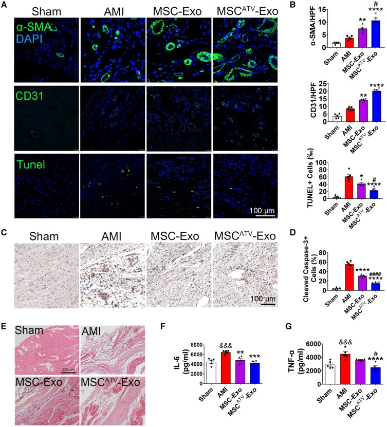
Figure 5.
MSCATV-Exo promoted angiogenesis and reduced cardiac apoptosis and inflammation after infarction. (A) Neovascularization at the border zone on Day 28 post-MI was identified by staining with α-SMA and CD31 (green) and nuclei (blue). TUNEL staining at the border zone on Day 28 post-MI with TUNEL (green) and nuclei (blue). Scale bar =100 μm. (B) Quantification of α-SMA+ cells, CD31+ cells and TUNEL+ cells in A (n = 5). (C) Cleaved Caspase-3 staining at the border zone on Day 28 post-MI. Scale bar =100 μm. (D) Quantification of Cleaved Caspase-3+ cells in C (n = 5). Apoptosis rate was quantified as the percentage of cells that were positive for TUNEL and Cleaved Caspase-3 staining. (E) HE staining at the border zone on Day 28 after MI. Scale bar =200 μm. (F and G) Quantification of IL-6 and TNF-α expression level in the infarct border zone tissue of rat heats using ELISA method (n = 5). All data are mean ± SEM. Statistical analysis was performed with one-way ANOVA followed by Tukey’s test. && P < 0.01, &&& P < 0.001 vs. Sham or Control group; *P < 0.05, **P < 0.01, ***P < 0.001, ****P < 0.0001 vs. AMI group; # P < 0.05, ## P < 0.01, ### P < 0.001, #### P < 0.0001 vs. MSC-Exo group.

