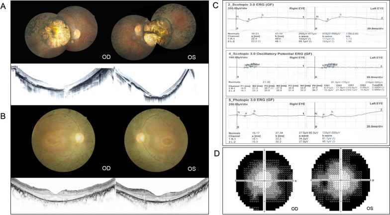
Fig. 1.
Clinical manifestations of typical and atypical RP patients. A Fundus photos of F2-III:3 presented bilateral prominent macular atrophy, with chorioretinal attenuation and extensive bone spicule pigmentation. OCT showed severe thinning of macular structure along with loss of photoreceptors. B–D Clinical manifestations of F5-III:1. B Fundus photos showed typical RP presentations, including waxy pallor disc, attenuated retinal vasculature, and mid-peripheral bone-spicule pigmentation. OCT illustrated severe loss of photoreceptors with preservation in the fovea. C Full-field electroretinogram (ERG) demonstrated a reduced rod and cone response amplitude. D Constricted visual field

