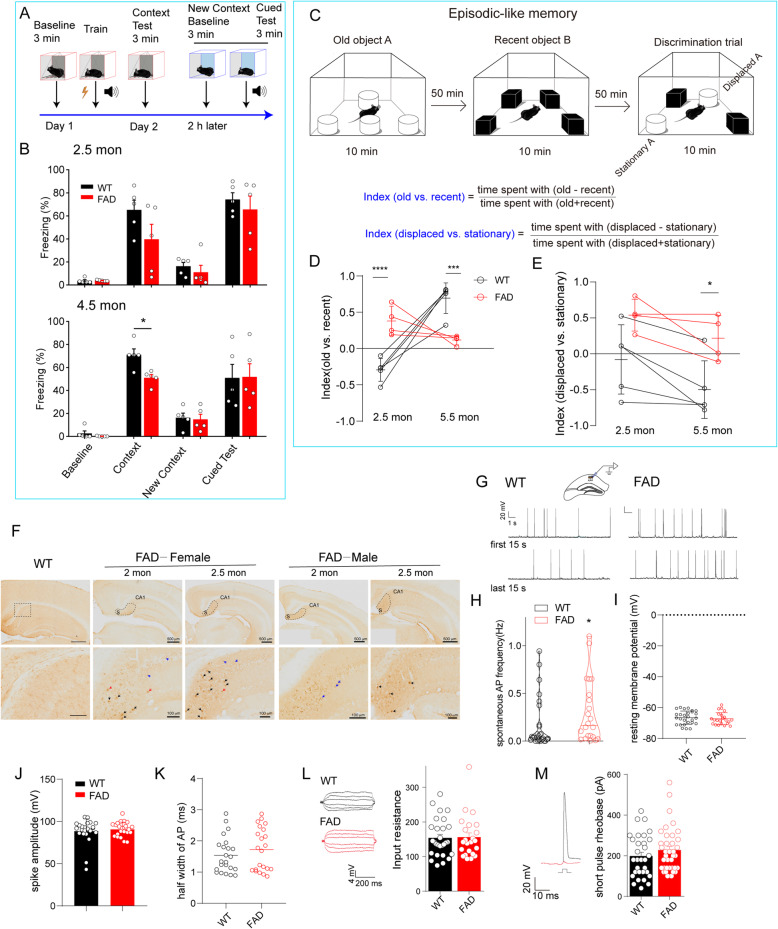
Fig. 1.
Hippocampal CA1 pyramidal neurons exhibited hyperactivity accompanied with few extracellular Aβ plaques in 5XFAD mice with mild cognitive decline. A Schematic diagram of contextual fear conditioning (CFC). B 5XFAD mice showed a significant reduction in freezing time at 4.5 months old (51.08 ± 2.76% vs. 71.14 ± 5.052%) while remained comparable level with WT mice at 2.5 months old (39.62 ± 29.4% vs. 65.19 ± 18.88%) in context test. Data are analyzed by two-way ANOVA with Bonferroni’s multiple comparisons test, mean ± SEM, *p < 0.05 vs. WT, n = 5 mice per group. C novel object recognition paradigm reflecting episodic-like memory. D, E The index (old vs. recent) indicating the ability to identify objects (D) and the index (displaced vs. stationary) indicating episodic memory of mouse (E) were subjected to statistical analysis. *p < 0.05, ***p < 0.001, ****p < 0.0001 vs. WT with two-way ANOVA, followed by Sidak’s multiple comparisons test, n = 5 in WT, n = 4 in FAD. Interaction (age × genotype) is significant (***p = 0.0002) in D and not significant in E. F Intraneuronal Aβ (stained with 6E10 antibody) signals were found in the cortex and hippocampus. The subiculum areas were surrounded by dashed lines (above), bar scale 500 μm. Obvious Aβ deposition was shown in a locally enlarged image of the subiculum (bottom) in a 2–2.5-month-old 5XFAD mouse. Red arrow: primary plaques; black arrow: canonical plaques; blue arrow: intraneuronal Aβ deposition. Bar scale 100 μm. G Action potential of the CA1 pyramidal neuron in WT (left) or 5XFAD mice (right) (female at 2.5 months old) was recorded with whole-cell current-clamp recording. The traces show the first 15 s and the last 15 s in at least 5 min recording time. H–K The spontaneous action potential (sAP) events during recording time (H), the resting membrane potential (I), the amplitude of spike (J), and the half-width of AP were detected automatically with Clampfit10.4. Unpaired Student’s t-test was used to compare these two groups, and the Mann–Whitney test was used in H, *p = 0.0380, n = 27 neurons/13 mice for WT; n = 22 neurons/10 mice for FAD, effect size is 0.299. In J and K, unpaired t-test with Welch’s correction was applied. Except for H, all values are presented as mean ± SEM. L Subthreshold stimulation was constituted of current injections from − 30 to + 30 pA in a duration of 500 ms, and the input resistance of CA1 pyramidal neurons in WT and 5XFAD mice was determined by Ohm’s law. M Step current injections (increment 10 pA) crossing the threshold were performed to determine the short-pulse rheobase of neurons. The red trace indicates the last subthreshold voltage response, and the black trace depicts the first firing triggered by suprathreshold current injection. The dots indicate individual neurons recorded, n = 28 (FAD), n = 23 (WT) in L; n = 31 (WT), n = 37 (FAD) in M. Unpaired Student’s t-test was used in L and M

