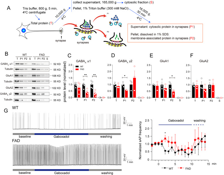
Fig. 4.
The attenuation of GABAA receptor sensitivity to agonist was accompanied with declined GABAA subunits localization in the synapses of the 5XFAD hippocampus. A Diagram for preparation of synaptic fractions. B Representative bands of western blotting for fractions from the WT and 5XFAD mouse hippocampus. C–F Normalized protein levels of GABAA α1 subunit (C), GABAA γ2 subunit (D), GluA1 (E), and GluA2 (F) distributed in each fraction (T, P1, P2, S, respectively) were subjected to unpaired Student’s t-test, *p < 0.05, **p < 0.01 vs. WT. The dots show the number of mice in each group, n = 5 mice/group in C, n = 3 mice/group in D–F. All values are presented as mean ± SEM. G GABAA receptor agonist, gaboxadol (GBX), in the final concentration of 5 μM was added in extracellular ACSF solution during slice whole-cell recording. The spontaneous action potential (sAP) of CA1 pyramidal neurons in WT or 5XFAD mouse slices was recorded, and the frequency was normalized to baseline. Values were analyzed by unpaired Student’s t-test for each time point, n(WT) = 5, n(FAD) = 8, *p = 0.0289 for 2 min

