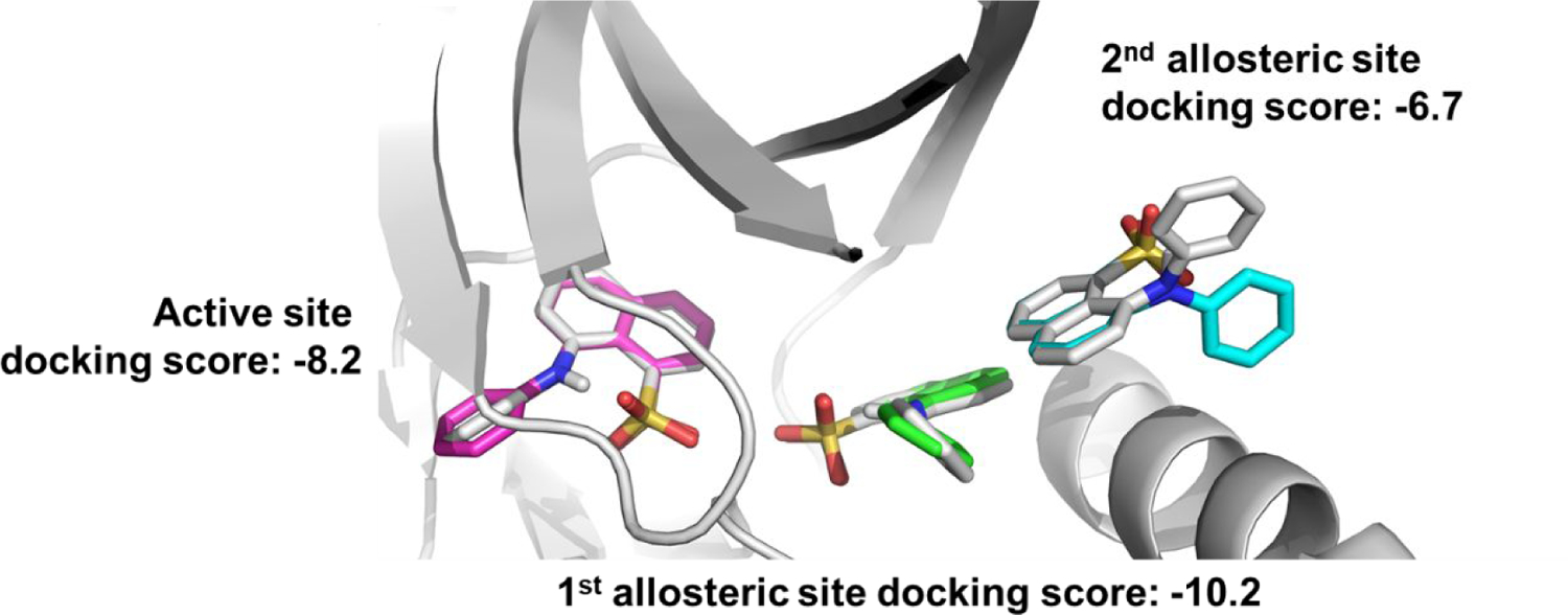
Figure 1.

Docking ANS molecules into co-crystal structure with Cdk2 (gray, PDB ID: 3PXQ (18)). At high concentrations of ANS, 3 molecules of ANS are bound – two in allosteric sites (labeled 1st allosteric site and 2nd allosteric site) and another in the active site. The top docking scores and binding poses of each ANS molecule into its respective site (active site = magenta, 1st allosteric site = green, 2nd allosteric site = cyan) predicts different binding affinities of ANS for each site.
