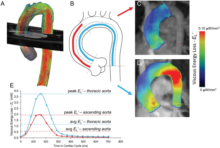
Figure 1:
(A) Aortic contour was segmented from the initial 4-dimensional-flow dataset with separation of the head and neck vessels. (B) A luminal centreline was then created to delineate 2 anatomically standardized aortic regions: (1) ascending aorta (sinotubular junction to the first arch vessel origin) and (2) thoracic aorta (sinotubular junction to mid-descending aorta measured at the level of the aortic root). Viscous kinetic energy loss represented by heat map visualization depicting the energy loss per voxel volume in the ascending aortic (C) and thoracic region (D).

