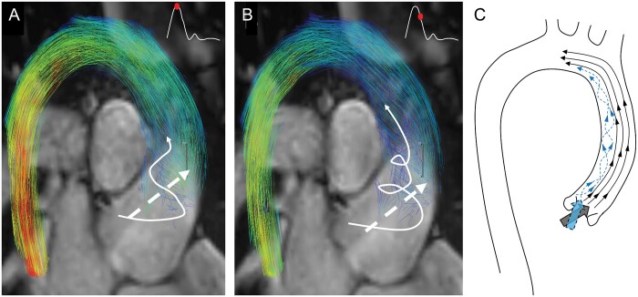
Figure 4:
Abnormal flow formations in a child with repaired tetralogy of Fallot (TOF) superimposed to standard steady-state free precession magnetic resonance imaging images for better visualization of the aortic outflow tract—flow relationship at the peak systole (A) and late systole (B). Characteristic secondary supraphysiological helices formed along the inner curve of the aorta and enlarged through the aortic lumen during progression of systole (white arrow). Secondary helical flow formations then mix with the primary flow jet (dashed white arrow) at the proximal level of the aortic arch. (C) Artistic representation of streamlines present in the thoracic aorta of patient with TOF. Two prominent stream jets at the level of the left ventricular outflow tract result in laminar cohesive flow along the outer curvature of the aorta, and secondary helical flow along the inner curve.

