
Figure 2.
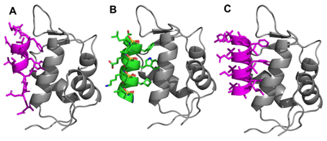
(A) Crystal structure of interaction of p53 with MDM2 (PDB: 1YCR). (B) Modeling of lead homogeneous l-sulfono-γ-AApeptide. (C) Designed homogeneous d-sulfono-γ-AApeptide 4 interaction with MDM2. P53 and homogeneous d-sulfono-γ-AApeptide are shown as magenta cartoon, homogeneous l-sulfono-γ-AApeptide is shown as green cartoon, and MDM2 is shown as gray cartoon.